

Cardiovascular system and artificial replacements
Research on tissues and body structures and biomaterials
Relationship between blood vessel wall architecture and constitutive behavior
Project description:
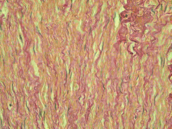
Histological section of venous wall. Stained with van Gieson. Red collagen fibrils are apparent.
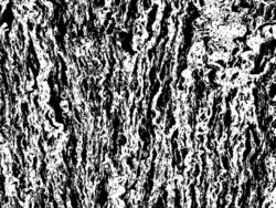
Collagen thrasholding (white color).
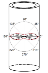
Resulting model of the distribution of collagen fibers orientations in venous wall.
Blood vessel wall is layered structure built with cellular and non-cellular components. The most important cells are endothelial cells and smooth muscles. Both may sense mechanical stress. It may lead to artery adaptation and remodeling in order to restore mechanical state. Smooth muscle cells create circular rings which are surrounded with collagen fibers (the most important load-carrying component). In medial layer, these musculo-fascial complexes are embedded in elastin membranes. The architecture and the arrangement are age and position dependent. The main aim is to find how such complicated structure determines physical phenomena observed within a loading. We use light microscopy methods and computer image analysis within blood vessel wall histomorphometry. Consecutively, this information is incorporated into constitutive models.
Hynek Chlup ; doc. Ing. Lukáš Horný Ph.D. ; Ing. Jakub Kronek Ph.D.Resesrchers:
Department of Forensic Medicine 3rd Faculty of Medicine CU, Institute of Thermomechanics AS CR, vvi
Cooperation:
Literature:
- Vesely, J., Horny, L., Chlup, H., & Zitny, R. (2011). Collagen orientation and waviness within the vein wall. Paper presented at the Computational Plasticity XI - Fundamentals and Applications, COMPLAS XI, 720-728.
- L. Horny, J. Kronek, H. Chlup, R. Zitny, J. Vesely and M. Hulan (2010) Orientations of collagen fibers in aortic histological section. Bulletin of Applied Mechanics, vol. 6, no. 22, p. 25-29
- L. Horny, J. Kronek, H. Chlup, R. Zitny and M. Hulan (2010) A distribution of collagen fiber orientations in aortic histological section. IFMBE Proceedings, vol. 29, p. 772-775
- L. Horny, M. Hulan, R. Zitny, H. Chlup, S. Konvickova and T. Adamek (2009) Computer-Aided Analysis of Arterial Wall Architecture. IFMBE Proceedings, vol. 25/4, p. 1494-1497