

Dental and orofacial system
Analysis of the loading influence TM joint on the other side owing to the implantation of total joint replacement
Project description:
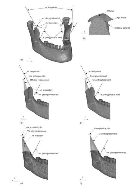
a) FE Model of the mandible, TM joint with articular fossa and masticatory muscles (anatomic configuration). Muscles (CONNECTOR elements) are represented by the lines. b) Detailed view of a cut in sagittal plane through mandible condyle, TM disc and fossa. c) A detailed picture of the Type A1 resection (only right part of the whole model is shown). d) A detailed picture of the Type A2 resection. e) A detailed picture of the Type B1 resection. f) A detailed picture of the Type A1 resection

The graphs show the relative FCP magnitude and its components [%] in the right and left TM joint for all resection types (relative i.e. values of the FCP for each type of resection compared to values of FCP obtained for anatomic configuration). The graph line on top represents the incisor bite; the graph line in the middle the molar bite and the graph line at the bottom represents the right molar bite (in this case, no results are available for a Type B2 resection). Total replacement of the TM joint is modeled on the right side
The temporomandibular (TM) joint is one of the most used joints in the human body. Any defect in this joint has a significant influence on quality of life. If the TM joint is damaged, treatment is very difficult and, in addition, it is impossible to replace full function of this joint by total replacement. The objective of this study was to create a parametric numerical FE analysis to compare the effect of surgical techniques used for total TM joint replacement implantation on loading the TM joint on the other side. Our hypothesis is that for the optimal function of all total TM joint replacements used in clinical practice is crucial minimally invasive surgical technique, when the minimum masticatory muscles have to be resected. This factor is more important than the design of the usually used total TM joint replacements. Range of the muscles resection influences the mechanical loading of the whole system. In the parametric FE analyses magnitude of TM joint loading was compared for four different ranges of muscle and bone resections (Fig. 1.) during bite to anatomical model. Results obtained from all FE analyses (Fig. 2.) support our hypothesis that increasing range of the muscles resection has a consequence of increased magnitude of the TM joint overloading the opposite side. In depend of the range of resection up to 235% for bite on incisor and up to 491% for bite on molars. Main conclusion from our study is recommendation to minimisation of muscle resection during replacement implantation and proposal to modification of the TM joint replacements condylar part attachment.
The results obtained from the numerical FE analyses of the effect of the implantation on the TM joint on the other side provide information for the design of a TM joint implant attachment which will enable smaller surgery in the system of masticatory muscles on the side of the implant. According to our FE analysis the design of the total replacement of mandible condyle and articulating fossa doesn't seem to be as big issue as the attachment of the condyle replacement to the mandible. Proper change in the attachment of the condyle replacement configuration of the total TM joint replacement can reduce the loading of the healthy TM joint on the other joint after implanting the total replacement. This will benefit the patient considerably. Suitable construction of a replacement attachment could mean that a total TM joint replacement will cease to be a treatment of last resort.
Resesrchers:
Stomatology clinic 1. LF a VFN (Radek Jirman, MD, MSc, MBA; Prof. Jiří Mazánek, MD, PhD)
Cooperation:
Literature:
- Jirman R., Horák Z., Bouda, T., Mazánek J., Řezníček J.: Influence of the method of TM joint total replacement implantation on the loading of the joint on the opposite side. Computer Methods in Biomechanics and Biomedical Engineering. Accepted in 2009
- Horák Z., Bouda T., Jirman R.: Loading of the temporomandibular joint after artificial joint implantation on the opposite side. In IV International Congress on Computational Bioengineering - Book of Abstract [CD-ROM]. Bologna: University of Bologna, 2009, 1, p. 91.
- Bouda T., Horák Z., Jirman R.: Identification of the Changes in Extent of Loading the TM Joint on the Other Side Owing to the Implantation of Total Joint Replacement. In Human Biomechanics 2008 International Conference, Extended Abstracts. Prague: Czech Technical University - Fac. of Mechanical Engineering, 2008, p. 11-12.
- Horák Z., Bouda T., Jirman R., Mazánek J., Řezníček J.: Identification of the Changes in Extent of Loading the TM Joint on the Other Side Owing to the Implantation of Total Joint Replacement. 2009, 13TH INTERNATIONAL CONFERENCE ON BIOMEDICAL ENGINEERING, VOLS 1-3, Book Series: IFMBE Proceedings, 23(1-3), 1535-1538